AI analysis software
for chest X-ray reading
Classifies 14 abnormal findings, localizes lesions and visualizes AI attention — all in one report.
01Overview
AI that supports the reader
TORAH Vision AI uses a ResNet50-based deep learning model to analyze high-resolution chest X-rays, presenting clinicians with the likelihood and location of abnormal findings. The clinician makes the final decision; the AI supports the process.
14 abnormal findings
From aortic enlargement to pneumothorax and pulmonary fibrosis — 14 findings and No Finding in a single analysis.
Grad-CAM visualization
Heatmaps overlaid on the original image show the regions the AI relied on.
Lesion localization
Per-finding bounding boxes indicate the location of suspected abnormalities.
High-resolution image processing
Uses the full grayscale depth of high-resolution medical images.
02Analysis Findings
14 abnormal findings + No Finding
The analysis classes defined in the product specification, grouped by anatomical region.
Descriptions are general radiological definitions provided for reference and do not represent the diagnostic performance of the product.
03Analysis Output
Actual analysis output
Review the original image, the Grad-CAM attention heatmap and the combined lesion-and-heatmap view side by side.
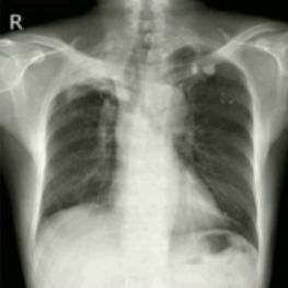
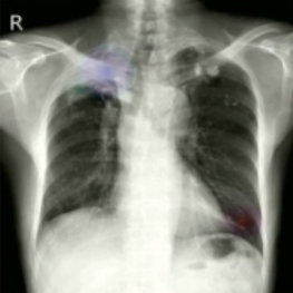
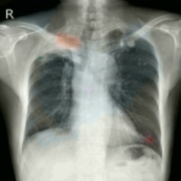
Excerpted from a TORAH Vision AI analysis report screen.
04Workflow
From image input to results
A five-step analysis process delivers results ready for clinical review.
-
STEP 01
Image input
Upload a chest X-ray in DICOM or another supported format.
-
STEP 02
Preprocessing
The high-resolution image is normalized and prepared for analysis.
-
STEP 03
AI analysis
The deep learning model analyzes the image and classifies findings.
-
STEP 04
Grad-CAM visualization
Regions the AI attended to are rendered as a heatmap.
-
STEP 05
Review
Review the report with per-finding scores and visualizations.
05Specifications
Product specifications
- Product
- TORAH Vision AI
- Type
- Medical AI software supporting chest X-ray reading
- Input
- Chest X-ray (DICOM, JPEG, PNG)
- Image processing
- High-resolution medical image processing
- Model
- ResNet50-based deep learning
- Analysis classes
- 14 abnormal findings + No Finding
- Output
- Per-finding scores, bounding boxes, Grad-CAM heatmap, integrated report
- Quality system
- ISO 13485, Digital Medical Device GMP
This product is software that supports clinicians in reading chest X-rays. The final clinical decision must always be made by a qualified clinician.
Contact
Let's talk about
medical AI for your institution
Product adoption, demo requests, clinical collaboration or international partnerships — our team responds within one business day.